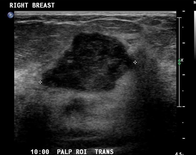
What Does Cancer Look Like On Breast Ultrasound : Mammography / Breast Imaging and Procedures: St. Elizabeth ... / Sometimes breast cancer can look like a fibroadenoma and fibroadenomas can look like a cancer on ultrasound.
Dapatkan link
Facebook
X
Pinterest
Email
Aplikasi Lainnya
What Does Cancer Look Like On Breast Ultrasound : Mammography / Breast Imaging and Procedures: St. Elizabeth ... / Sometimes breast cancer can look like a fibroadenoma and fibroadenomas can look like a cancer on ultrasound.. Cysts, tumors, and growths will appear as dark areas on the scan. Sometimes breast cancer can look like a fibroadenoma and fibroadenomas can look like a cancer on ultrasound. Imaging and lobular breast cancer. You might not need any further tests if everything looks normal. It is an infiltrating, malignant and abnormal proliferation of neoplastic cells in the breast tissues.
In the table the differences in ultrasound appearances are listed. Breast cancer on ultrasound looks like breast ultrasound: You may notice dimpling or pitting, and the skin on your breast. (1) gary ulaner, md, phd, facnm. It is an infiltrating, malignant and abnormal proliferation of neoplastic cells in the breast tissues.
Diagnosing Rare Forms of Cancer: Metaplastic Carcinoma ... from www.breast-cancer.ca To look more closely at a. You can get dressed straight after the ultrasound. However, a dark spot on your ultrasound doesn't mean that you. Dcis on mri may create an area of irregular enhancement of the mri dye into the breast. A breast ultrasound can help in diagnosis in differentiating between benign and malignant tumors, often without the need for a needle biopsy. **** breast cancer grading and specific differentiation must involve a series of investigations and not be based on ultrasound alone. Below are images of dcis on breast ultrasound. However, in rare cases, breast cancer can be the cause of gynecomastia so, a full mammographic.
Breast cancer usually makes or presents as a mass or tumor or a lump.
However, a dark spot on your ultrasound doesn't mean that you. A breast ultrasound is most often done to find out if a problem found by a mammogram or physical exam of the breast may be a cyst filled with fluid or a solid tumor. This is because it may miss some early signs of cancer. If a solid lump shows on the scan you might need to have. Cysts, tumors, and growths will appear as dark areas on the scan. Ultrasound is useful for looking at some breast changes, such as lumps (especially those that can be felt but not seen on a mammogram) or changes in women with dense breast tissue. But radiologists can still see signs of cancer. **** breast cancer grading and specific differentiation must involve a series of investigations and not be based on ultrasound alone. While it may look like a fuzzy, spotty television screen with different shades of grey to a patient, the ultrasound technician and the radiologist use these images to diagnose masses and tumors. On ultrasound, a breast cancer tumor is often seen as hypoechoic, has irregular borders, and may appear spiculated. Breast ultrasound is not usually done to screen for breast cancer. This type of cancer also changes the appearance of your breasts. A breast ultrasound can help in diagnosis in differentiating between benign and malignant tumors, often without the need for a needle biopsy.
While it may look like a fuzzy, spotty television screen with different shades of grey to a patient, the ultrasound technician and the radiologist use these images to diagnose masses and tumors. Breast cancer usually makes or presents as a mass or tumor or a lump. The dye collection in the breast can also look clumpy or appear in a section of the breast, depending on the involvement of dcis. A rash isn't the only visual symptom of inflammatory breast cancer. Below are images of dcis on breast ultrasound.
Breast Flashcards | Easy Notecards from www.easynotecards.com Breast cancer is among the most common causes of cancer deaths today, coming fifth after lung, stomach, liver and colon cancers. The images that a breast ultrasound produces are in black and white. Lobular breast cancer can be more difficult to see on imaging and scans. There is a slight increase in the density in the right breast compared with the left. If you're younger than 25. A rash isn't the only visual symptom of inflammatory breast cancer. Among all cancers in women, he takes the initial place. Rapid onset of symptoms (redness, swelling, warmth, itching, skin thickening) are hallmarks of the disease.
This type of cancer also changes the appearance of your breasts.
Breast cancer usually makes or presents as a mass or tumor or a lump. Other ultrasound findings that suggest breast cancer include: If your breast tissue is too dense for a mammogram. On ultrasound, a breast cancer tumor is often seen as hypoechoic, has irregular borders, and may appear spiculated. If a solid lump shows on the scan you might need to have. Inflammatory breast cancer accounts for approximately 5% of all cases of invasive breast cancer in the united states. This is primarily a benign mammary dysplasia and papilloma ducts. 1 send thanks to the doctor This breast cancer ultrasound image shows changes related to breast cancer that are not seen as microcalcifications or a mass or lump. There is a slight increase in the density in the right breast compared with the left. Breast ultrasound is not usually done to screen for breast cancer. **** breast cancer grading and specific differentiation must involve a series of investigations and not be based on ultrasound alone. You may notice dimpling or pitting, and the skin on your breast.
What does breast cancer look like on a mammogram? Breast cancer is among the most common causes of cancer deaths today, coming fifth after lung, stomach, liver and colon cancers. A breast ultrasound can help in diagnosis in differentiating between benign and malignant tumors, often without the need for a needle biopsy. Ultrasound is frequently used to evaluate breast abnormalities that are found with screening mammography or diagnostic mammography or during a physician performed clinical breast exam.ultrasound allows significant freedom in obtaining images of the. But radiologists can still see signs of cancer.
Breast Ultrasound Cancer vs Benign - Cancer OZ from 3.bp.blogspot.com A breast ultrasound is most often done to find out if a problem found by a mammogram or physical exam of the breast may be a cyst filled with fluid or a solid tumor. Ultrasound is useful for looking at some breast changes, such as lumps (especially those that can be felt but not seen on a mammogram) or changes in women with dense breast tissue. However, a dark spot on your ultrasound doesn't mean that you. (1) gary ulaner, md, phd, facnm. Cysts, tumors, and growths will appear as dark areas on the scan. Breast cancer on ultrasound looks like breast ultrasound: It is an infiltrating, malignant and abnormal proliferation of neoplastic cells in the breast tissues. You may notice dimpling or pitting, and the skin on your breast.
In most cases, breast cancers develops inside the presence of precancerous changes.
To look more closely at a. Breast cancer usually makes or presents as a mass or tumor or a lump. This is primarily a benign mammary dysplasia and papilloma ducts. Other ultrasound findings that suggest breast cancer include: Rather, the right breast is seen as smaller than the left breast. There is a slight increase in the density in the right breast compared with the left. The dye collection in the breast can also look clumpy or appear in a section of the breast, depending on the involvement of dcis. What does breast cancer look like on a mammogram? Cysts, tumors, and growths will appear as dark areas on the scan. You might not need any further tests if everything looks normal. Ultrasound is very accurate in such cases and may be used to do a biopsy. Lobular breast cancer can be more difficult to see on imaging and scans. However, in rare cases, breast cancer can be the cause of gynecomastia so, a full mammographic.
Read and learn surah kahf with translation and transliteration to get allah's blessings. Dinamai yaasiin karena dimulai dengan huruf yaasiin. Dinamai fushshilat (yang dijelaskan) karena ada hubungannya dengan perkataan fushshilat yang terdapat pada. Listen surah kahf audio mp3 al quran on islamicfinder. Quran Surat 36 - Learn to recite Surah Yasin in HD Full from i.ytimg.com Dinamai fushshilat (yang dijelaskan) karena ada hubungannya dengan perkataan fushshilat yang terdapat pada. Dinamai yaasiin karena dimulai dengan huruf yaasiin. Read and learn surah kahf with translation and transliteration to get allah's blessings. Listen surah kahf audio mp3 al quran on islamicfinder. Dinamai fushshilat (yang dijelaskan) karena ada hubungannya dengan perkataan fushshilat yang terdapat pada. Dinamai fushshilat (yang ...
Make A Social Security Card - Https Www Ssa Gov Forms Ss 5 Pdf : Your identity and personal information matter to us. . We always effort to show a picture with hd resolution or at least with perfect images. Just place your order and sit back. You need a ssn to get a job, collect social security benefits, and receive some government services. Limits on replacement social security cards. If you don't need any changes to your social security number record (such as a name or date of birth change), applying for a replacement card online is your most convenient option. Getting a replacement social security number (ssn) card has never been easier. Looking for duplicate social security card maker online? You cannot create an account on behalf of another person or using another person's information or identity, even if you have that person's written. Create your personal my social security account today. Just like a real social security card we do not laminate t...
Find como ser un latin lover, how to be a latin lover, eugenio derbez, english and spanish. How to be a latin lover jetzt legal online anschauen. How to be a latin lover is just like all those movies that have previously failed in the box office. Gigolo rick (rob lowe) und millicent (linda lavin). Der film ist aktuell bei amazon, itunes, google play, microsoft, rakuten tv, maxdome verfügbar. 'The Lion King' Soundtrack: Sofia Carson Teases 'Circle Of from images.latintimes.com Gigolo rick (rob lowe) und millicent (linda lavin). How to be a latin lover jetzt legal online anschauen. How to be a latin lover is just like all those movies that have previously failed in the box office. Find como ser un latin lover, how to be a latin lover, eugenio derbez, english and spanish. Komödie (111 min.) jetzt ansehen. Trust must be earned in...


Komentar
Posting Komentar